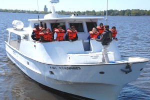
Falls Lake Partners in Forensic Science Program 2024
2024 Falls Lake Partners in Forensic Science Application-
only for MSMMS, EGMMS and CCMMS students
The Center for Applied Aquatic Ecology
 The main mission of this center is to provide service to faculty and graduate students across NCSU, and service to the people of North Carolina, through our State-certified water quality laboratory, our phytoplankton microscopy laboratory, our real-time remote monitoring systems that serve as early warning systems to help safeguard major drinking source waters, our long-term, high-frequency datasets on selected surface waters, and our extensive outreach education Floating Classroom Program for secondary and high school students. A second goal is to provide water quality research information needed to augment regulatory and non-regulatory programs aimed at optimizing management and conservation of water resources, through (i) provision of training and support opportunities for advanced undergraduate students, graduate students, and post-doctoral fellows; (ii) relevant applied research on freshwater, estuarine and marine resources of the State, with emphasis on chronic and acute impacts of nutrient over-enrichment and related pollution on harmful algal blooms; and (iii) development of information databases that contribute to environmental education and education outreach about the interdependence between ecosystem functions and natural resource management.
Learn more about what we do below!